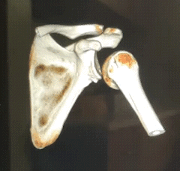
Shoulder Dislocations: Reduction and Stabilization
Epidemiology
The shoulder is the most commonly dislocated joint, making up nearly 50% of major joint dislocations. In the United States, the incidence of shoulder dislocations is approximately 23.9 per 100,000 person years. Anterior shoulder dislocations account for up to 98% of cases, posterior shoulder dislocations account for 2-4%, inferior, superior, and intrathoracic dislocations make up <1%. Shoulder dislocations tend to occur more often in young males (age 20-30 years) with male to female ratio of 9:1, and in older females (age 61-80 years) with female to male ratio of 3:1.
How does it happen?
Specific mechanisms of injury or elements of history may suggest certain types of shoulder dislocation. For example, traumatic injury to the abducted, externally rotated, and extended arm often occurring during sports is associated with anterior shoulder dislocation; seizures and lightning/electrical injuries are associated with posterior dislocation; axial load to an extremely abducted arm is associated with inferior dislocation. Possible associated injuries include humerus fracture, rotator cuff tear, axillary/radial nerve injury, arterial injury, Bankart lesion, Hill-Sachs lesion, among others.
Patient Presentation
Patients with acute shoulder dislocation will typically present with shoulder pain, inability to move the shoulder, and may have signs of neurovascular injury. In cases of anterior dislocation, the arm is held in slight abduction and external rotation, will have a “squared off” appearance to the deltoid, humeral head may be palpable anteriorly, and the patient will resist internal rotation.
In cases of posterior dislocation, the arm is held in adduction and internal rotation, anterior shoulder may appear flat with prominent coracoid process, palpable humeral head beneath the acromion posteriorly, and the patient will resist external rotation.
In cases of inferior dislocation, the arm is often fully abducted with the elbow flexed behind the patient’s head, and the humeral head may be palpable along the lateral chest wall.
In all patients regardless of type of dislocation, it is important to assess for neurologic and vascular injuries by checking distal pulses, and assessing axillary and radial nerve function.
Workup
Diagnostic imaging includes plain shoulder radiographs to confirm diagnosis and identify any associated pathology such as fracture.
Routine pre- and post-reduction films include AP view, scapular Y view, and axillary view. For patients with recurrent dislocations, prereduction films may not be necessary. Administer analgesics to decrease pain and allow the patient to assume a position of comfort. Obtain informed consent for the reduction procedure and gather necessary materials. Of note, orthopedic surgery should be consulted for patients with concomitant fracture and elderly patients due to higher risk of complications during reduction.
Treatment/Management
There are a variety of treatment approaches including procedural sedation, intra-articular lidocaine, US-guided brachial plexus or interscalene block, and a plethora of reduction techniques. Generally, the reduction method depends on clinician preference and the patient’s condition. Some notable techniques include scapular manipulation, external rotation, Milch technique, Stimson technique, traction-countertraction, Spaso technique, Davos technique, among others.
After successful reduction, confirmation with post-reduction radiograph, and ensuring the patient remains neurovascularly intact, it is important to immobilize the shoulder in a position of stability. This is especially important to reduce risk of further injury and recurrent dislocation. For anterior shoulder dislocations, this is typically achieved by having the patient in a position of adduction and internal rotation using either a collar and cuff, sling and swathe, or shoulder immobilizer.
However, for patients with posterior dislocation placing them in a shoulder immobilizer would put them in a position of instability (internal rotation and adduction); therefore, it is important to have these patients stabilized in external rotation or in abduction with neutral rotation. This can be achieved utilizing commercial orthotic braces such as an ultrasling or gunslinger brace.
The length of time for immobilization varies depending on type of injury and patient age; thus, patients should follow-up with an orthopedic surgeon within 5-7 days and should remain immobilized until they are evaluated by an orthopedist in clinic.
Final Thoughts
Although the vast majority of shoulder dislocations treated in the ED will be anterior, it is imperative to be aware of differences in managing posterior dislocations. Putting the patient in a position of instability will significantly increase their risk of recurrent dislocation and return to the ED; therefore, posterior dislocations should be stabilized with a brace in external rotation or abduction. Give your friendly neighborhood orthotist a call!
Michael Simoes, MD
USF Emergency Medicine
Michael Simoes is a current PGY-1 Emergency Medicine resident at USF. He completed his medical school education at Florida Atlantic University. His academic interests include simulation, EMS, and global health.
References
Sharon R Wilson, MD, Trevor John Mills, MD, MPH. Shoulder Dislocation in Emergency Medicine. (2018) https://emedicine.medscape.com/article/823843-overview
Scott C Sherman, MD, Allan B Wolfson, MD, Jonathan Grayzel, MD, FAAEM. Shoulder Dislocation and Reduction. (2020) https://www.uptodate.com/contents/shoulder-dislocation-and-reduction
Zacchilli MA, Owens BD. Epidemiology of shoulder dislocations presenting to emergency departments in the United States. J Bone Joint Surg Am. 2010 Mar;92(3):542-9. doi: 10.2106/JBJS.I.00450. PMID: 20194311.
Pescatore R, Nyce A. Managing Shoulder Injuries in the Emergency Department: Fracture, Dislocation, and Overuse. Emerg Med Pract. 2018 Jun. 20 (6):1-28
Robinson CM. The epidemiology, risk of recurrence, and functional outcome after an acute traumatic posterior dislocation of the shoulder. J Bone Joint Surg Am. 2011;93(17):1605–1613.
Images obtained from orthobullets.com, uptodate.com, and shoulderelbow.org; CT reconstruction of posterior shoulder dislocation obtained from patient encounter at Tampa General Hospital.


